In this article, you will get information about why you need a wrist MRI, Wrist MRI scan center in delhi, Anatomy and wrist MRI price in delhi, etc.
Wrist pain, whether mild or severe, can significantly affect an individual’s quality of life, limiting daily activities and productivity. Magnetic Resonance Imaging (MRI) plays a pivotal role in assessing wrist pain by providing detailed insights into the underlying causes and determining the extent of discomfort. This information is crucial for deciding whether conservative or invasive treatment is necessary. Furthermore, MRI findings are instrumental in planning the surgical approach for treating wrist conditions.
Table of Contents
Why Is Wrist MRI Needed?
Wrist MRI is needed for several reasons:
- Detailed Imaging: MRI provides a comprehensive view of bones, ligaments, tendons, cartilage, and nerves, making it the gold standard for diagnosing complex wrist issues.
- Unclear Diagnosis: When X-rays or clinical assessments fail to pinpoint the cause of wrist pain, MRI offers detailed visualization of abnormalities.
- Surgical Planning: For patients requiring surgery, MRI helps surgeons plan the procedure by accurately identifying the extent and location of the issue.
- Early Detection: MRI can detect subtle changes in bone and soft tissue structures, aiding in the early diagnosis of conditions like Kienböck’s disease or ligament tears.
- Non-Invasive: As a non-invasive diagnostic tool, MRI eliminates the need for exploratory surgery in many cases.
Importance of Wrist MRI in Diagnosis and Treatment
Clinical assessments sometimes fail to identify the exact cause of wrist pain. In such cases, wrist MRI offers a computational method for accurate diagnosis, enabling healthcare professionals to deliver suitable patient care. The imaging results help surgeons decide on the appropriate intervention, whether it is arthroscopic surgery, open surgery, or a combination of both. Understanding whether the origin of wrist discomfort is intra-articular (within the joint) or extra-articular (outside the joint) is critical for effective treatment planning.
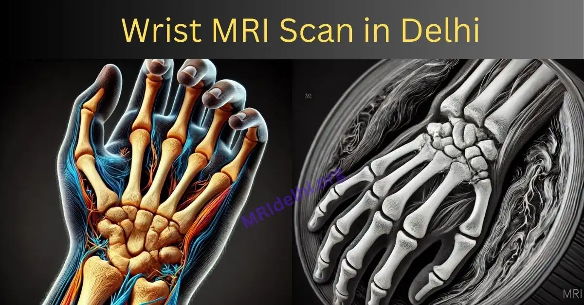
Anatomy of the Wrist in MRI
The wrist is a complex joint composed of:
- Carpal Bones: Eight small bones arranged in two rows that form the wrist joint.
- Ligaments: These structures connect the carpal bones to each other and to the forearm bones (radius and ulna), providing stability.
- Tendons: Tendons in the wrist allow movement by connecting muscles to bones.
- Nerves and Blood Vessels: The median, ulnar, and radial nerves, along with associated blood vessels, pass through the wrist, supplying sensation and blood flow to the hand.
MRI captures detailed images of these structures in three planes—coronal, axial, and sagittal—allowing for accurate assessment of any abnormalities.
Intra-Articular Wrist Pain
Intra-articular pain originates from issues within the wrist joint itself and can be categorized into disorders involving bone or soft tissue. Common intra-articular bone conditions include:
- Fractures: MRI can detect subtle fractures that may not be visible on X-rays.
- Avascular Necrosis (AVN): This condition involves the death of bone tissue due to a lack of blood supply, commonly affecting the lunate bone (Kienböck’s disease).
- Styloid-Carpal Impaction Syndrome: Caused by abnormal contact between the radial styloid process and the scaphoid bone.
- Ulnar-Carpal Impaction Syndrome: A degenerative condition caused by the ulnar head impinging on the lunate bone.
Kienböck’s Disease
Several classification systems, including the Lichtman categorization, have been employed to evaluate Kienböck’s disease. However, arthroscopic classification systems are now preferred by many surgeons for their practicality in surgical planning. Familiarity with these systems enhances the radiologist’s ability to contribute to surgical decision-making and ensures better outcomes for patients.
Extra-Articular Wrist Pain
Extra-articular wrist pain arises from structures outside the joint. Common causes include:
- Tendon and Tendon Sheath Injuries: Conditions such as tenosynovitis or tendon tears can be effectively evaluated using MRI.
- Neuromas: Benign growths of nerve tissue that may cause localized pain.
- Sheath Tumors: Tumors within the nerve sheaths, such as ganglion cysts or other benign soft tissue masses.
- Accessory Muscles: Variations in muscle anatomy that may lead to compressive symptoms.
Optimizing Wrist MRI Imaging
MRI imaging of the wrist requires acquisition in three planes: coronal, axial, and sagittal. Each plane provides unique and complementary information:
- Axial Images: Excellent for visualizing soft tissues, blood vessels, nerves, and the radiocarpal articulations.
- Coronal Views: Best suited for examining ligaments and bones.
- Sagittal Views: Useful for evaluating the wrist’s overall structural alignment and detecting abnormalities in soft tissue and bone.
High-resolution imaging protocols should include sequences such as T1-weighted, T2-weighted, and STIR (Short Tau Inversion Recovery) to enhance visualization of different tissue types.
Wrist MRI Cost in delhi
The cost of a wrist MRI can vary based on factors such as location, imaging facility, and whether contrast is required. On average:
- Wrist MRI Without Contrast price in delhi : The cost typically ranges from Rs. 2,599 to Rs. 5,999
- Wrist MRI With Contrast price in delhi : Adding contrast can increase the cost from Rs. 7,000 to Rs. 8,999.
- Insurance Coverage: Many insurance plans cover MRI scans, but it is essential to confirm coverage details beforehand.
- Additional Fees: Some facilities may charge extra for radiologist interpretation or same-day reports.
Frequently Asked Questions (FAQs)
-
Why is an MRI recommended for wrist pain?
MRI provides detailed images of bones, soft tissues, ligaments, and nerves, enabling accurate diagnosis of conditions causing wrist pain. It is especially useful when X-rays or physical examinations fail to identify the cause.
-
How does an MRI help in surgical planning?
MRI findings help surgeons determine the nature and extent of wrist pathology, allowing them to decide on the most appropriate surgical approach, whether arthroscopic or open.
-
Can MRI detect early stages of Kienböck’s disease?
Yes, MRI is highly sensitive in detecting early changes in bone structure and blood supply, making it the preferred modality for diagnosing Kienböck’s disease.
-
How long does a wrist MRI take?
A typical wrist MRI takes about 20-30 minutes, depending on the imaging protocol and the need for contrast enhancement.
-
Are there any risks associated with wrist MRI?
MRI is a safe procedure without ionizing radiation. However, patients with certain implants or devices (e.g., pacemakers) should inform their doctor beforehand.
Conclusion
Wrist MRI is an indispensable tool in diagnosing and managing wrist pain. By providing a detailed view of both intra-articular and extra-articular structures, MRI helps clinicians make informed decisions about treatment strategies. Its role in surgical planning is particularly significant, ensuring precise interventions and improved patient outcomes. As imaging technology continues to advance, the utility of wrist MRI will only expand, solidifying its place in modern medical diagnostics and care.


1 thought on “Wrist MRI in Delhi”